


About the Cover
Vol. 98 No. 5 (2022)
Fertilization is the starting point to create a new life in sexually reproducing organisms. At this time, the highly differentiated oocyte and sperm fuse with each other by fertilization and are converted into a pluripotent early embryo. In this process, the cytoplasmic components derived from each gamete are selectively degraded as the embryos start zygotic expression to change their properties toward early development. Recent studies have revealed that lysosomal degradation systems such as endocytosis and autophagy also play pivotal roles in the remodeling of intracellular components for early development.
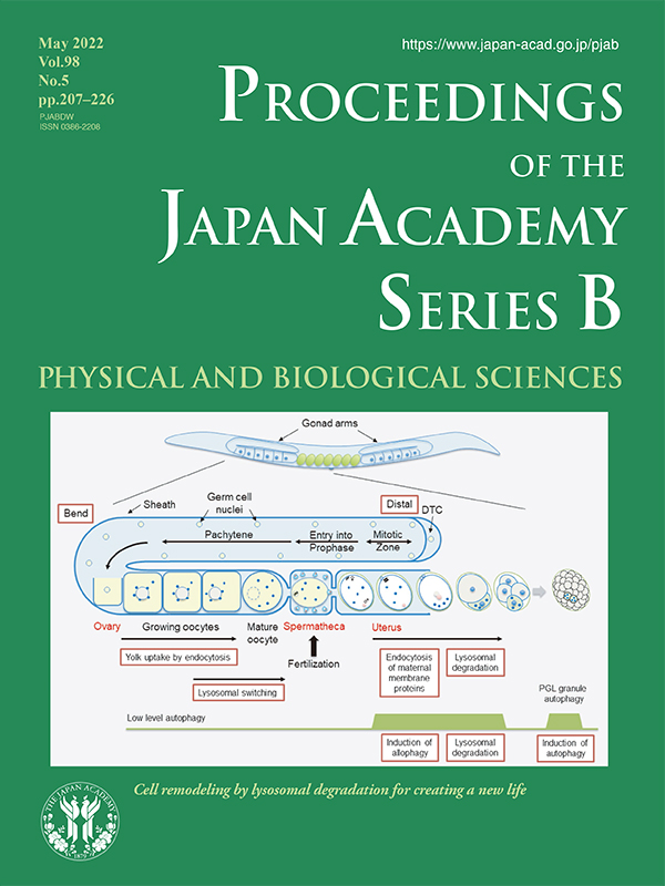
The nematode Caenorhabditis elegans is hermaphroditic and generally reproduces by self-fertilization. A C. elegans adult hermaphrodite contains two U-shaped gonads connecting to a spermatheca that accommodates sperms. Oocytes are formed by cellularization around the bend region of the gonadal arm and growing oocytes move to the proximal region. In the most proximal region, oocytes receive a signal from the sperm and undergo meiotic maturation and cortical rearrangement. Mature oocytes are ovulated to the spermatheca containing sperm and fertilized. The fertilized egg then moves to the uterus, completes meiosis I and II, and starts embryogenesis.
In the gonad, growing oocytes take up yolk components via receptor-mediated endocytosis. Before fertilization, oocytes undergo lysosomal switching that converts the pH of the lysosomal lumen from non-acidic to acidic and upregulates lysosomal functions in response to sperm signaling. Sato (see the review article in this issue, pp. 207-221) and colleagues discovered that sperm-derived paternal mitochondria and membranous organelles are ubiquitinated at metaphase I immediately after fertilization, and then eliminated by allogenic (nonself) organelle autophagy, named allophagy during embryogenesis. This selective degradation of paternal mitochondria by autophagy ensures maternal inheritance of mitochondrial DNA in C. elegans. They also revealed that some populations of maternal membrane proteins are ubiquitinated at anaphase II and selectively endocytosed for degradation by the 2-cell stage. Induction of autophagy occurs again at later embryonic stages (64-100 cells). P granules are germ cell lineage-specific non-membranous ribonucleoprotein granules. After fertilization, P granules are gradually formed during the 1-cell stage and specifically distributed to the germ cell lineage during embryogenesis. When P granule components are distributed to somatic cells of embryos, they are removed by selective autophagy. In autophagy-deficient mutant embryos, P granule components form aberrant PGL granules in somatic cells.
Asako Sugimoto
Professor, Graduate School of Life Sciences, Tohoku University