


About the Cover
Vol. 97 No. 8 (2021)
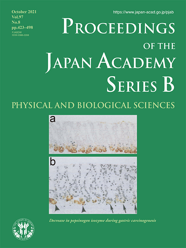
Decrease in pepsinogen isozyme during gastric carcinogenesis
Gastric cancer remains one of the most common and fatal cancers worldwide, especially among older men. Gastric cancer prevention has been a long-standing challenge. An experimental gastric carcinogenesis model using N-methyl-N´-nitro-N-nitrosoguanidine (MNNG), a known mutagen, was developed by Sugimura and Fujimura in 1967. Around 1970, it was postulated that gastric cancer was induced by chemical carcinogens in food or in the environment or by chemical substances produced in the body. Sugimura’s colleagues, Furihata and Tatematsu et al., demonstrated that decreased or absent expression of a pepsinogen isozyme, PG1, was induced in the gastric mucosa and gastric adenocarcinomas after administration of MNNG to rats through analysis using polyacrylamide gel electrophoresis. In this figure (see the review article in this issue (pp. 462-478)), normal pyloric mucosa without carcinogenic potential (a. normal control) and normal-looking pyloric mucosa within it (b. + MNNG) are well contrasted. PG1 in pyloric gland cells of rat stomach was demonstrated immunohistochemically, stained with anti-rat PG1 serum and proliferative cell kinetics were analyzed using immunohistochemical staining for bromodeoxyuridine (BrdU) incorporation. Control normal pyloric gland cells were stained in brown and cells in the proliferative zone, confined to the region of the isthmus, were labelled with BrdU (a). Arrows indicate pyloric gland cells with decreased PG1 plus BrdU labeling, and arrowheads indicate pyloric gland cells with high PG1 content located in the lower one-third of the pyloric gland without BrdU labeling after continuous BrdU administration (b). The life span of cells comprising glands with decreased PG1 was estimated to be ~6-8 days, whereas that of cells in normal pyloric glands was ~11-13 days. Thus, the data indicate that pyloric gland cells with decreased PG1 demonstrated a degree of independence from surrounding pyloric glands with regard to proliferation kinetics, suggesting that pyloric glands with decreased PG1 are a preneoplastic lesion involved in gastric carcinogenesis. It was demonstrated that altered DNA methylation affects PG1 gene expression in rat glandular stomach mucosa after MNNG treatment. These findings led to human PGI and PGII studies, and decreases in PGI in the human gastric mucosa and serum were identified as markers of atrophic gastritis. In the 1990s, other researchers revealed that Helicobacter pylori bacteria causes atrophic gastritis and later gastric cancer. In the 2000s, a gastric cancer risk screening method combining assays to detect serum anti-H. pylori IgG antibodies and serum PGI and PGII levels, the “ABC method”, was established by Miki et al. Eradication of H. pylori and endoscopic follow-up examination after the ABC method are recommended to prevent gastric cancer and detect it in earlier stage for better treatment outcomes.
Mitsuhiro Fujishiro
Professor, Department of Gastroenterology, Graduate School of Medicine, The University of Tokyo