


About the Cover
Vol. 94 No. 9 (2018)
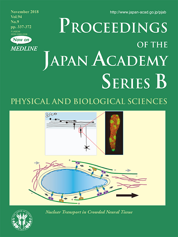
Cell migration is critical for various physiological and pathological processes. During development of the mammalian brain, newborn neurons migrate from their birthplaces to final destinations to form ordered structures such as layers (where neurons are laminated) or brain nuclei (where neurons group to form clusters). The upper left diagram exemplifies granule cells migrating (initially horizontally then vertically) in the early postnatal cerebellar cortex, which is highly crowded with numerous cells (not illustrated for simplicity). A key step in neuronal migration is the delivery of the nucleus, which packages DNAs densely and is the largest cargo in each cell, within such crowded neural tissues, where interstitial spaces that would freely permit the entrance of new cells are limited.
The upper right photomicrograph shows that the nucleus, stained with H3K9Me3 (green) and H4K20Me2/3 (red), adopts a unique shape in a vertically (inward) migrating cerebellar granule cell, which appears to be appropriate for moving against physical resistance (similar to the first car of a Shinkansen bullet train). In this issue (pp. 337-349), Kengaku reviews, with a thought-provoking bird’s-eye view and deep molecular insights, recent findings on the mechanisms of force transmission by cytoskeletal motors and actomyosin during nuclear migration. Various model cells, including mesenchymal or fibroblastic cells, myoblasts, Caenorhabditis elegans hypodermal cells, and Drosophila melanogaster oocytes, are compared with neurons in the developing mammalian brain. This review shed light on tremendous efforts (labor) of neurons to build our brains.
The lower diagram illustrates the nuclear migration mechanisms revealed by Kengaku and colleagues through high-resolution confocal imaging. The perinuclear microtubules (green lines with plus [+] and minus [-] ends to both directions) dynamically bind to small points on the nuclear envelope via kinesin (orange wheels; plus-oriented motor) and cytoplasmic dynein (purple wheels; minus-oriented motor). These bidirectional motors induce sharpening, rotation, and translocation of the nucleus independently of the force produced by F-actin (magenta lines), and thereby squeeze the nucleus through narrow interstitial spaces in the developing brain.
Takaki Miyata
Professor, Department of Anatomy and Cell Biology
Nagoya University Graduate School of Medicine