


About the Cover
Vol. 94 No. 3 (2018)
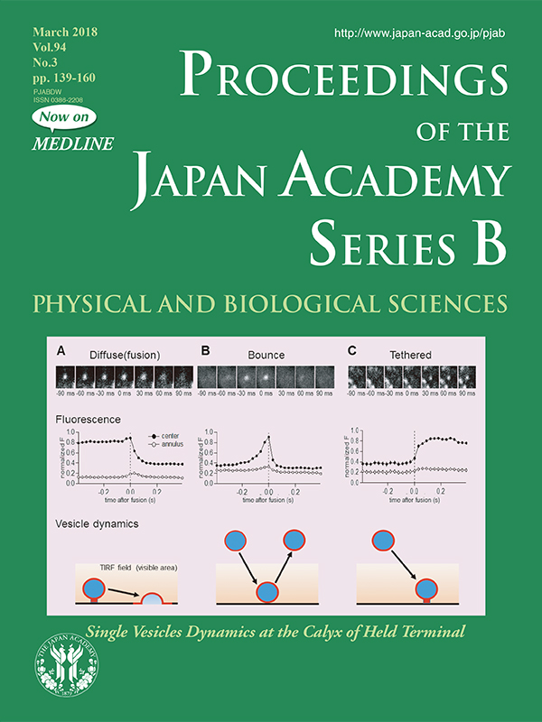
In the nerve terminal, neurotransmitters are filled at high concentrations in intracellular organelles called synaptic vesicles, and are released into the extracellular space by exocytosis. Presynaptic action potentials synchronize exocytosis of multiple vesicles, but it remains unknown how stimulation regulates the movements of these vesicles. In the present review (pp. 139-152), Takeshi Sakaba introduces new findings from his lab on vesicle behaviors near the terminal membrane in response to stimulation. Vesicles, loaded with fluorescent probe, were monitored in real-time using TIRF microscopy in acutely dissociated nerve terminals of the calyx of Held. TIRF imaging is characterized by its high spatio-temporal resolution and restricted optical field within 100 nm from a membrane in contact with the bottom of a chamber. These optical properties enable the visualization of vesicle movements across the optical border and the exocytotic release of fluorescent probe from vesicles. Before stimulation (time 0 indicated by dashed lines in the Fluorescence panels), many vesicles remained within the optical field (A, C), but some vesicles moved into the field, as revealed by an increase in fluorescence intensity (B). Upon stimulation, some vesicles started to lose their fluorescent probe by diffusion, with a 100-ms order time constant, indicating exocytosis (“Fusion”, A). Vesicles that moved into the field (B) often moved out upon stimulation, as deduced from a relatively fast decline in fluorescence intensity (“Bounce” in B). Some other vesicles moved into the field upon stimulation (C) and remained there, presumably by anchoring to the terminal membrane (“Tethered”).
Upper panels of the figure are reproduced from figure 2 of Midorikawa and Sakaba (2015) Neuron 88, 492-498.
Distinguished Professor
Okinawa Institute of Science and Technology